By Dr. Mark S. Siegel
A picture of the retina may one day help diagnose people with Alzheimer’s before they show any symptoms.
A study published in “Investigative Ophthalmology and Visual Science” showed that the technique could identify Alzheimer’s in mice. The procedure will be tested in humans next.
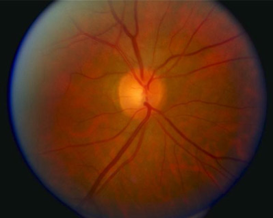
Researchers at the University of Minnesota Center for Drug Design were able to identify Alzheimer’s from detailed color images of the retinas of mice.
The retina is made from similar kinds of tissue as the brain and that tissue can be seen directly by looking into the eye. As Alzheimer’s begins, it changes the way that light is reflected off of the retina. The study showed that in mice this change starts long before any behavioral or memory changes are noticeable.
Alzheimer’s causes changes in memory, thinking and vision, as well as movement and behavior problems. The symptoms become more severe over the course of the disease.
According to the Alzheimer’s Association, the disease is caused by the buildup of amyloid plaques and tangles in the nerve cells in the brain. These plaques are difficult to see in living brains, so Alzheimer’s is usually only diagnosed from the symptoms.
By the time symptoms become obvious, a person will already have lost some brain function.
There is no treatment for the buildup in the brain that causes Alzheimer’s. But there are treatments for some of the symptoms. There are also treatments that can slow the disease’s progress.
If this new test works as well in humans as it did in mice, people with Alzheimer’s could begin treatment to slow down the disease years earlier, before any symptoms are seen.
Dr. Mark S. Siegel is the Medical Director at Sea Island Ophthalmology on Ribaut Road in Beaufort.


